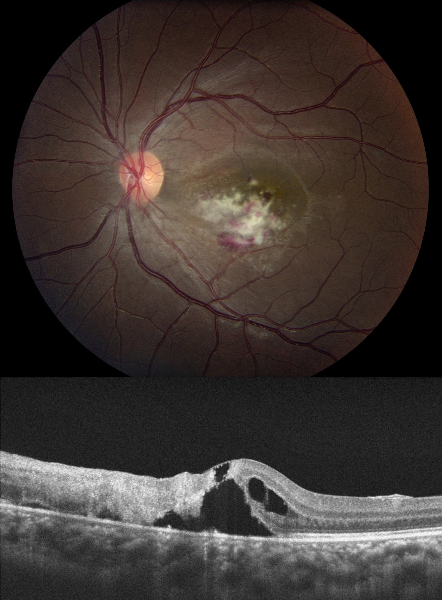

In the active phase, it occurs as necrotizing chorioretinitis with overlying vitritis. In 70 to 80% of cases, it can occur as a unilateral focal chorioretinal lesion. In the inactive stage, it can present as quiescent atrophic chorioretinal scars, which appear as pigmented lesions in clusters. Typical features are excavated macular lesions with associated pigmentation.Īcquired Toxoplasmosis: Most infections are thought to occur congenitally and can remain asymptomatic for several years and can become clinically evident commonly in the second through fourth decades. Congenital toxoplasmosis is associated with retinochoroidal lesions in 80% of infected newborns. The presence of chorioretinitis, intracranial calcifications, and hydrocephalus are considered the classic triad of congenital toxoplasmosis. However, fetuses infected in early pregnancy are more likely to show clinical signs of infection. In the first trimester rate of transmission to the fetus is around 10 to 25%, in the third trimester the transplacental transmission is approximately 60 to 80%. The severity of fetal involvement is related to the gestational period at which infection occurred, with more severe in early trimesters, when fetal death may result. It can cause severe infection in immunosuppressed individuals and pregnant women.Ĭongenital Toxoplasmosis: In congenital toxoplasmosis, the mother is often asymptomatic or has mild constitutional manifestations. Infection usually occurs by either consumption of tissue cysts present in raw or undercooked meat or by ingesting oocysts in the feces of cats. Toxoplasma gondii is the most common cause of infectious posterior uveitis worldwide.

The extent of ocular involvement varies depending on the organism. Since the choroid is responsible for the vascular support of the outer layers of the retina, inflammation of these layers can lead to vision-threatening complications.Ĭhorioretinitis is usually due to an infectious etiology. The choroid is the vascular layer of the eye, which lies between the retina and the sclera. Chorioretinitis is a type of posterior uveitis. Uveitis also classifies as granulomatous or nongranulomatous, acute, chronic or recurrent, insidious, or sudden, based on criteria like appearance, duration, or type of onset. The Standardization of Uveitis Nomenclature (SUN) consensus conference workshop classified uveitis into anterior (iritis, iridocyclitis, anterior cyclitis), intermediate (pars planitis, posterior cyclitis, hyalitis), posterior uveitis (choroiditis, chorioretinitis, retinochoroiditis, retinitis, and neuroretinitis) and panuveitis based on the anatomical location. Though the term uveitis means inflammation of the uveal tract (iris, ciliary body, and choroid), it can involve the adjacent structures like the retina, retinal vessels, vitreous, optic nerve head, and sclera. Chorioretinitis is a type of uveitis involving the posterior segment of the eye, which includes inflammation of the choroid and the retina of the eye.


 0 kommentar(er)
